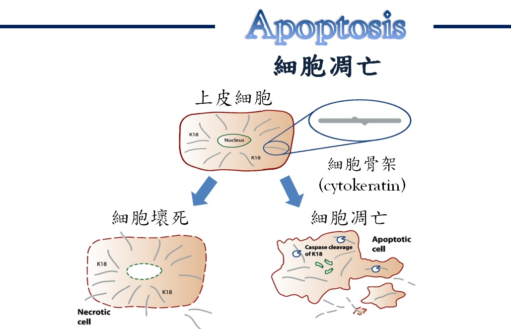
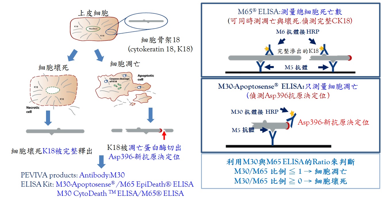
Apoptosis是一種細胞受環境刺激後,在基因調控之下所產生的自然死亡現象,故亦稱之為細胞程式死亡,與necrosis(細胞壞死)有所區別。Necrosis一般被認為是細胞在遭受物理性傷害後所造成的惡性死亡,其特徵包括胞器遭受嚴重破壞以及細胞膜喪失其完整性等,然而這些現象都不是在基因調控之下所產生的。而在發生apoptosis的細胞中,可發現細胞質中產生小泡囊、染色質濃縮以及DNA斷裂等現象。 Apoptosis與necrosis二者在in vivo主要的差別是在於前者不會引起發炎反應。總而言之,apoptosis是一種對周圍細胞及組織傷害最小的一種細胞自殺性死亡。
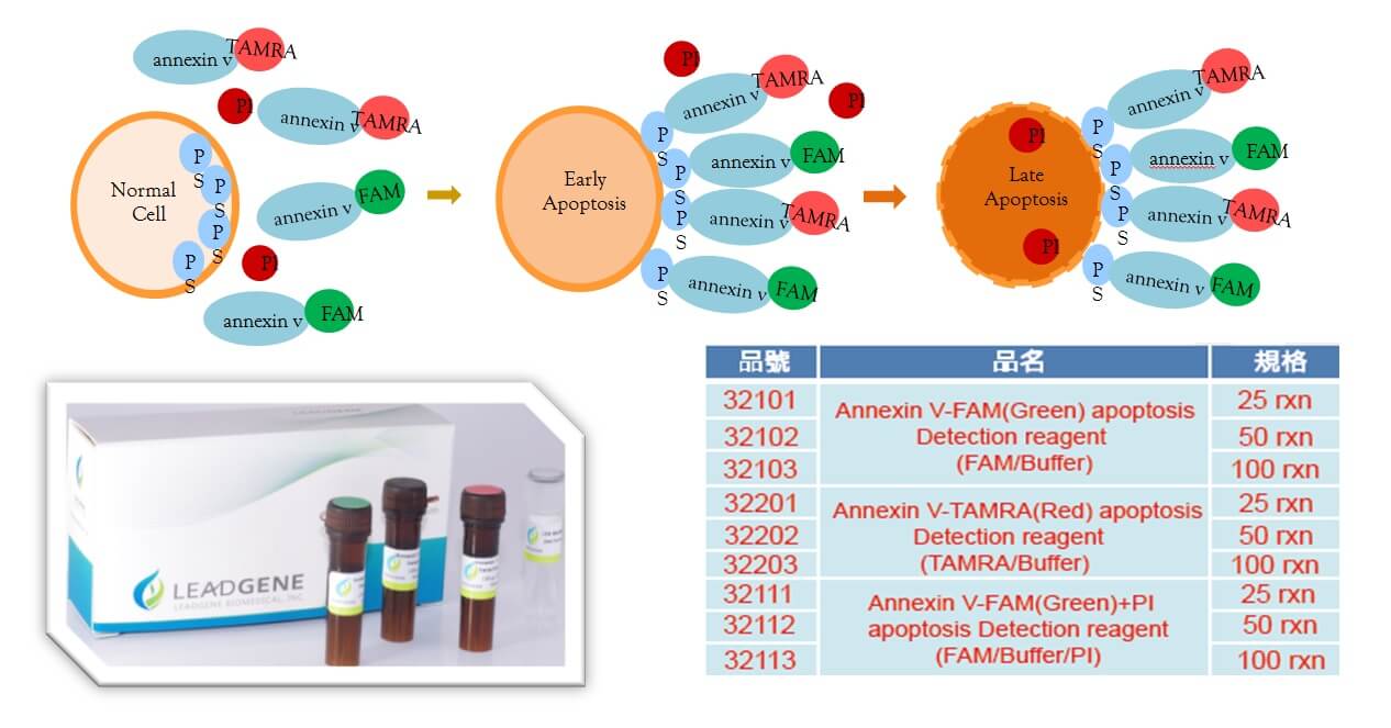
Apoptosis早期,Phosphatidyl-serine(PS)會由細胞膜內外翻到膜外表現,可利用AnnexinV,以流式細胞儀的方式做第一階段的偵測。
Apoptosis過程中,caspase的活化是引發細胞凋亡的共同特徵。活化的caspase會進行一連串的蛋白質酶解作用,因此本公司為研究者準備一系列的caspase antibodies,給您更得心應手的工具。可運用於WB、IHC、IF、FC。
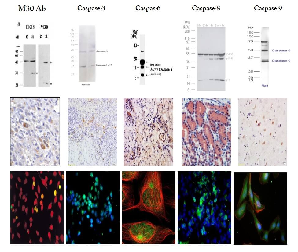
| M30 | caspase3 | caspase6 | caspase8 | caspase9 |
| 10700 | bs-0081R | bs-0151R | bs-0052R | bs-0049R |
Apoptosis最後,活化的caspase瀑布會引發DNA的片段化以及凋亡蛋白酶的裂解效應(Cleavage of Caspase Substrate),一般都以TUNEL assay作為是否走向apoptosis的最後證據,但是!!!DNA的斷裂或片斷化由於檢體的處理跟檢測方式的選擇,TUNEL assay的結果變得不是那樣的絕對!
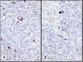
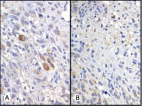
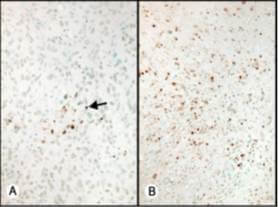
A: 細胞凋亡
B: 壞死細胞
從TUNEL ASSAY裡可觀察出細胞凋亡但同時壞死的腫瘤裡也會觀察到DNA斷裂的現象,比較活化的Caspase-3與M30出現細胞凋亡的一致性。
針對上皮細胞骨架CK18被caspase酶切出新的抗原決定位做為檢測標的。
關鍵字: M5、M6、M30、M65、M30/M65 ratio、上皮細胞、cCK18。

可運用方法學:
酵素免疫分析法 (ELISA)
西方墨點法 (Western blot)
流式細胞術 (Flow cytometry)
免疫細胞化學法 (ICC)
免疫組織化學法 (IHC)
可使用檢體:
-
細胞
-
組織
-
血清或血漿
-
培養過細胞的上清液
細胞凋亡檢測新工具 “細胞骨架CK18”
適用各種不同方法學及樣本,檢測細胞死亡準確,操作方便!!!
臨床運用範圍:
-
腫瘤治療評估
-
器官移植物排斥(GvHD)
-
肝炎治療評估(NASH.NAFLD.HBV.HCV)
-
其他器官損傷之評估,(自體免疫性疾病…等)
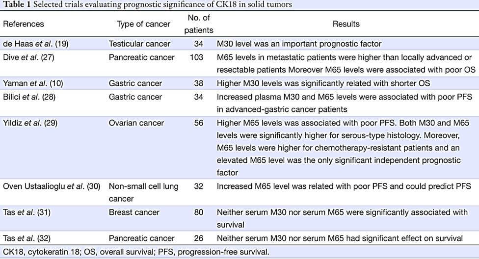
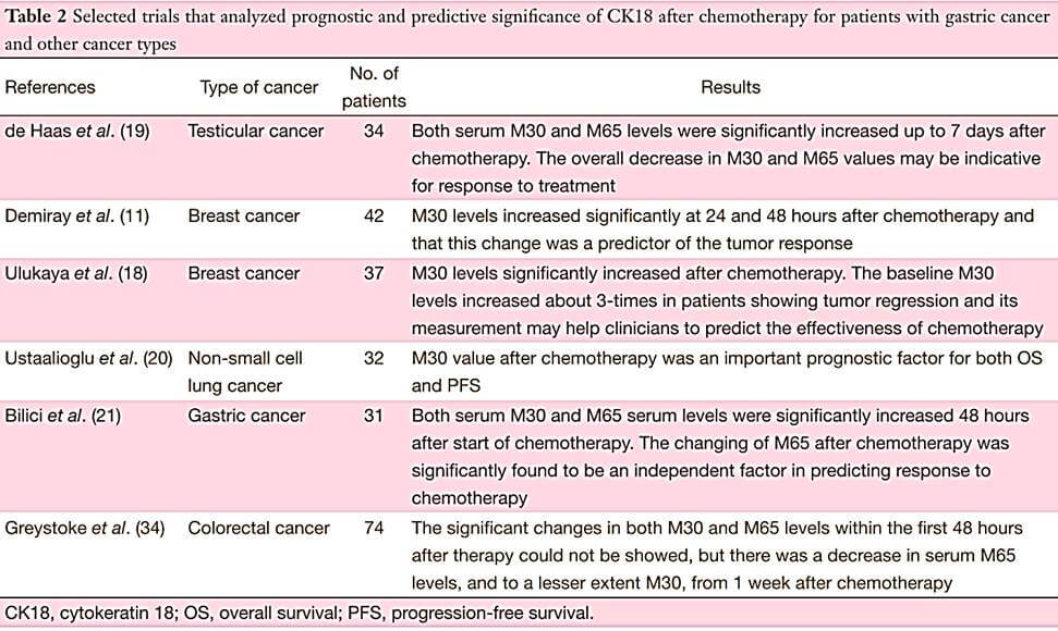
CK18於癌症化療病人意義之文獻:Cytokeratin 18 for chemotherapy efficacy in gastric cancer
CK18 may modulate intracellular signaling and apoptosis via interactions with various related proteins. There is evidence to show that CK18 is involved in the invasive or growth properties of tumors. Caspase-cleaved CK18 (M30) and total CK18 (M65) are measured in the circulation by enzyme-linked immunoabsorbent assays (ELISA). The measurement of M30 or M65 from epithelial-derived tumors could be a simple, noninvasive way to monitor or predict tumor progression and prognosis. However, there are conflicting results in vitro and in vivo. These discrepancies may be due to differences among the carcinoma types, therefore the precise roles of CK18 are currently unknown. But, particularly, in patients with gastric cancer, testicular cancer and colorectal cancer, serum M30 and/or M65 levels could be used as biomarkers to evaluate treatment response and they might guide in determining of the most appropriate combination chemotherapy regimen.
